Canalicular Lacerations


Overview

 
Canalicular lacerations are breaks (interruptions) in the normal tear duct drainage system. If not repaired promptly, tearing will usually result.

This systems originates with the puncta (there is one in both the upper and the lower eyelid) and is a conduit for tears to travel from the eyelid through the nasolacrimal sac into the nose.

Loading image. Please wait
"Tear system. a = lacrimal gland b = superior lacrimal punctum c = superior lacrimal canal d = lacrimal sac e = inferior lacrimal punctum f = inferior lacrimal canal g = nasolacrimal canal"
"Medial wall of left orbit."
"Lacrimal bone visible near center"
"Bone: Lacrimal bone"
"G_005_16"
"G_005_01"
"Gray1199"
"Gray894"
"Alveoli of lacrimal gland"
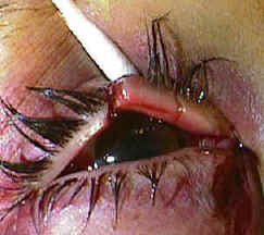
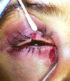
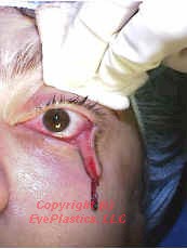
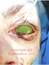
"The lacrimal apparatus is the physiologic system containing the orbital structures for tear production and drainage"
"The tarsal glands, etc., seen from the inner surface of the eyelids"
"Sympathetic connections of the sphenopalatine and superior cervical ganglia"
"Nerves of the orbit. Seen from above."
"The ophthalmic artery and its branches"


Tension, from trauma such as a blow from the fist, can result in an eyelid laceration which involves the canalicular system.

Repair requires re-approximation of the eyelid as well as re-approximation of the conduit; this is best achieved with a stent such as with silastic and fine sutures such as 6,7, or 8-0 vicryls..


The photos below show a patient who was hit in their right eye with a fist and who sustained a canalicular laceration:


This photo below shows a patient who was hit in their right eye with a fist and who sustained a canalicular laceration. This photo below shows a patient who was hit in their right eye with a fist and who sustained a canalicular laceration.

Treatment

There are several different means to repair such an injury. Placement of a stent (silastic tubing) helps maintain proper alignment of the conduit and prevent stricture after the repair.

Mono-canalicular stent Mono-canalicular stent Mono-canalicular stent
  • Following dilation and preliminary probing of lacrimal ducts, the Ritleng Probe (S1-1460u) is introduced into the canaliculus and nasolacrimal duct until contact is made with the nasal fossa floor.

  • The probe is pulled back slightly (1 cm) to facilitate the introduction of the prolene thread-guide into the nasal cavity.

  • The probe is oriented with its slit side facing anteriorly and pushed backwards so that the inferior end of the probe is facing anterior, thus
    directing the prolene towards the front of the nasal cavity.

  • The prolene is threaded through the probe to obtain a large loop which spreads out in the nasal cavity making it easy to locate. Retrieval of the blue prolene is easy when it appears in the anterior portion of the nose.

  • The prolene is retrieved under nasal illumination and visual control (nasal endoscope) with endonasal forceps or with the Ritleng Hook (S1-1480u)

Ritleng mono-canalicular stent
  • If the prolene thread-guide is not easily located in the anterior portion of the nose, or if it takes a posterior direction, the following technique is used for retrieval:

    • The probe is introduced until contact is made with the nasal fossa floor.

    • Metal-to-metal contact is made using the Ritleng Hook (S2-1480u) high up in the inferior meatus near the exit of the nasolacrimal duct.

  • The probe is then rotated 180 degrees while keeping the metal-to-metal contact with the hook thus orienting its inferior opening towards the back.

  • The hook should be above the probe's opening and the prolene.

  • This will enable the hook to catch the prolene loop when removing from the nose.

Ritleng mono-canalicular stent
  • The probe is slowly backed out of the inferior meatus and as soon as the metal-to-metal contact between the probe and the hook is lost, the hook catches the prolene loop and is carefully removed from the nose.

  • The probe is removed from the canaliculus and detached from the stent by sliding the thinner light blue portion of the prolene out through the probe's slit.

  • The prolene thread-guide is pulled out the nose along with the attached silicone tubing.

  • This same technique is used to intubate the second canaliculus in the case of a bicanaliqilar intubation.
    In the case of a monocanalicular intubation, the punctal plug at the other end of the silicone tubing is seated in the punctum using a punctal plug dilator inserter (S1-3090u).

These two photos show a canalicular laceration and its repair with a monocanalicular stent
using the .
his photo shows a canalicular laceration and its repair with a monocanalicular stent using the Ritleng probe.

Watch the a movie using the Ritleng Silicone Intubation system

References

  1. Conlon MR, Smith KO, Cadera W, Shun D,Allen LH. An animal model studying construction techniques and histopathologic changes in repair of canalicular a lacerations. Can J Ophthalmol 1994;29:3d
  2. .Kennedy RH May J, Dailey J, Flanagan JC Canalicular laceration: an 11 year epidemiologic and clinical study. Ophthalmic Plastic and Reconstructive Surg
  3. Loft HJ.Wobig JL. Dailey RA. The bubble test: an atraumatic method for canalicular laceration repair. Ophthalmic Piastic ann Reconstructive Surg 1996;12:61-64.
  4. Long JA. A method of monocanalicular silicone intubation. Ophthalmic Surg 1988 19 204 205
  5. McLeish WM Bowman B, Anderson RL The pigtail probe protected by silicone intubation a combined approach to cana icu ar reconstruction. Ophthalmic Surg 1992;23:281-283.
  6. Reifler DM. Management of canalicular laceration. Surv Ophthalmol 199136:11 j132
  7. Ritleng, Peirre. A simplifed technique for lacrimal intubation. Ocular surgery news. Vol 14, No 7