Basal Cell Carcinoma of the Eyelid
The most common eyelid cancer — its pearly, telangiectatic appearance, and Mohs/excisional treatment with oculoplastic reconstruction.
Medically reviewed by EyePlastics Medical Editorial BoardASOPRS oculoplastic surgeonsLast updated June 2026
Part of our complete guide to Eyelid Skin Tumors — this page covers basal cell carcinoma in depth.
Basal Cell Carcinoma
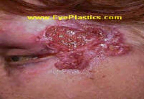
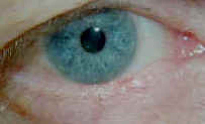
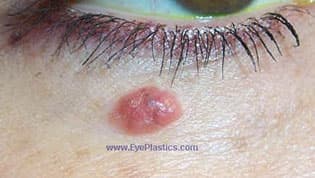
What Basal Cell Carcinoma Looks Like
A pearly or waxy nodule with rolled borders and fine surface vessels; some erode into a non-healing ulcer.

Basal cell carcinoma (BCC) is the most common eyelid malignancy, accounting for approximately 90% of all eyelid cancers. It arises from basal keratinocytes of the epidermis and is strongly associated with cumulative UV exposure, fair skin, and immunosuppression. The lower eyelid accounts for ~50% of cases, followed by the medial canthus (~25%), upper eyelid (~15%), and lateral canthus (~10%).
Clinical subtypes:
- Nodular BCC (most common) — pearly or translucent nodule with telangiectatic vessels and a rolled, indurated border; may ulcerate centrally (“rodent ulcer”)
- Morpheaform (sclerosing) BCC — flat, waxy, scar-like plaque with ill-defined margins; most aggressive subtype, frequently underestimated clinically; requires widest surgical margins
- Superficial BCC — flat, erythematous plaque; more common on the trunk; less frequent on the eyelid
Warning Signs — When to See a Specialist
Have any eyelid lesion evaluated promptly if it:
- Is new, growing, or changing in size, shape, or color
- Bleeds, crusts, or fails to heal
- Has a pearly or translucent surface with fine surface blood vessels
- Distorts the lid margin or causes loss of eyelashes (madarosis)
- Recurs after a previous removal
Lash loss and lid-margin distortion are especially important — they point toward a malignant rather than a benign process.
Surgical management: Mohs micrographic surgery with same-day oculoplastic reconstruction is the gold standard for periocular BCC. Mohs achieves the highest cure rate (5-year recurrence <1% for primary BCC) with the greatest tissue preservation — critical in the eyelid where even a few millimeters matter for function. For nodular BCC of the lower lid not involving the lid margin, wide local excision with frozen-section margin control is an alternative.
BCC is locally destructive but rarely metastasizes (<0.1%). Orbital invasion, though uncommon, can occur with neglected medial canthal BCC and may require orbital exenteration. Hedgehog pathway inhibitors (vismodegib, sonidegib) are used for locally advanced or metastatic BCC not amenable to surgery.
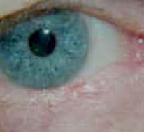
What Basal Cell Carcinoma Looks Like
Basal cell carcinoma classically appears as a pearly nodule with rolled borders and fine surface vessels, often with central ulceration and loss of lashes when the lid margin is involved. The lower eyelid and medial canthus are the most common locations.

Frequently Asked Questions
- How is eyelid basal cell carcinoma treated?
- Typically by complete removal (often Mohs micrographic surgery for clear margins) followed by oculoplastic reconstruction of the eyelid to restore function and appearance.
- Is basal cell carcinoma dangerous?
- Basal cell carcinoma rarely spreads to distant sites, but on the eyelid it can invade locally and damage the eye and orbit if untreated — so timely removal is important.
- Is basal cell carcinoma of the eyelid dangerous?
- It rarely spreads to other parts of the body, but on the eyelid it can grow into surrounding tissue and, if neglected, into the eye socket. Removed early, the cure rate is very high.
- What is Mohs surgery and why is it used on the eyelid?
- Mohs micrographic surgery removes the cancer in thin layers, checking each under the microscope until the margins are clear. It gives the highest cure rate while sparing the most healthy tissue — essential on the eyelid, where every millimeter matters for function.
- Will my eyelid look and work normally after removal?
- In most cases, yes. An oculoplastic surgeon reconstructs the eyelid in the same setting, restoring the lid margin, lash line, and tear drainage so the eye stays protected and the appearance looks natural.
Find a Specialist
Connect with a board-certified oculoplastic surgeon who specializes in basal cell carcinoma of the eyelid.
Search the Directory →