Watery Eye & the Evaluation of Tearing
Why the eye waters (epiphora) and how an oculoplastic surgeon evaluates it — history, clinical tests, irrigation, and imaging of the tear-drainage system.
Medically reviewed by EyePlastics Medical Editorial BoardASOPRS oculoplastic surgeonsLast updated June 2026
Part of our complete guide to Tear-Duct & Lacrimal Surgery — this page covers the watery-eye work-up in depth.
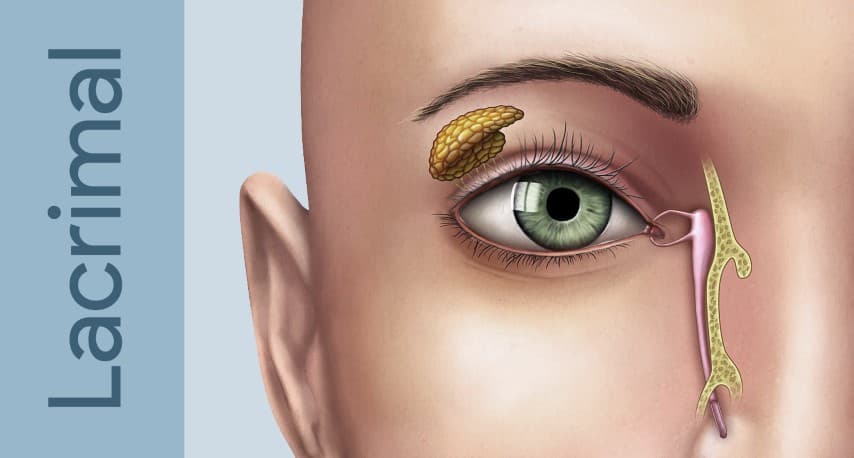
Evaluation of Tearing
A systematic evaluation identifies the site and severity of obstruction before planning treatment.
History
- Duration and laterality of tearing
- Associated discharge, pain, or swelling over the lacrimal sac
- Prior eye surgery, chemotherapy, or topical medication use (especially anti-glaucoma drops)
- History of facial trauma, nasal surgery, or chronic sinusitis
- Prior episodes of dacryocystitis
Clinical Tests
- Dye disappearance test (DDT): fluorescein dye placed in both eyes; persistence of dye on slit lamp after 5 minutes indicates delayed drainage. Quantified by asymmetry between eyes
- Jones I test (primary dye test): cotton swab placed beneath the inferior turbinate; recovery of fluorescein confirms functional patency of the entire system
- Jones II test (secondary dye test): if Jones I is negative, the sac is irrigated with clear saline; recovery of fluorescein-stained fluid from the nose indicates dye reached the sac (functional or partial obstruction at/below the sac), whereas recovery of clear saline only suggests dye never reached the sac (punctal/canalicular dysfunction)
- Lacrimal irrigation / probing: a fine cannula irrigates the system through the punctum. Hard stop (probe reaches the medial wall of the sac against the lacrimal bone) indicates a patent canalicular system up to the sac, localizing any obstruction at or below the sac; soft stop (probe meets spongy resistance before reaching bone) suggests canalicular obstruction proximal to the sac. Reflux of fluid indicates nasolacrimal obstruction
- Regurgitation test: pressure over the lacrimal sac expresses mucoid or purulent material through the punctum — confirms an obstructed, infected sac (dacryocystitis)
Imaging
- CT scan of orbits and sinuses: identifies bony anatomy, nasolacrimal canal dimensions, and sinus pathology; essential before revision DCR surgery
- Dacryocystography (DCG): contrast injected into the system outlines the anatomy and identifies the level of obstruction
- Nasal endoscopy: evaluates the nasal cavity, inferior meatus, and valve of Hasner; identifies intranasal pathology (polyps, deviated septum) that may contribute to obstruction
Frequently Asked Questions
- Why does my eye water constantly?
- A constantly watering eye (epiphora) usually means tears are either overproduced (often from irritation or dry eye) or, more commonly, not draining — because the tear-drainage pathway is narrowed or blocked.
- How is the cause of a watery eye diagnosed?
- Through a focused history and exam, the dye-disappearance test, and gentle irrigation/probing of the tear ducts; imaging is added when the level of blockage needs to be confirmed.
Find a Specialist
Connect with a board-certified oculoplastic surgeon who specializes in watery eye & the evaluation of tearing.
Search the Directory →