Treatment of blocked tear ducts, chronic tearing, dacryocystorhinostomy (DCR), and lacrimal infections — adult and pediatric.
Medically reviewed by EyePlastics Medical Editorial BoardASOPRS oculoplastic surgeonsLast updated June 2026
The lacrimal system is the eye’s tear apparatus — the glands that produce tears and the channels that drain them away. With every blink, a thin tear film is spread across the surface of the eye, keeping it lubricated, optically clear, and protected against infection; the used tears then drain through tiny openings at the inner corner of the eyelids, down through the tear sac and nasolacrimal duct, and into the nose. Because this single system both makes and removes tears, the problems that arise fall into two broad groups: insufficient tears or an unstable tear film, which leaves the eye dry, gritty, and irritated, and impaired drainage, which causes a constantly watering eye (epiphora), mucous discharge, and recurrent infection of the tear sac (dacryocystitis). The sections below follow the tears along this pathway — from how they drain, through the relevant anatomy, to the evaluation and surgical treatment of blockage.
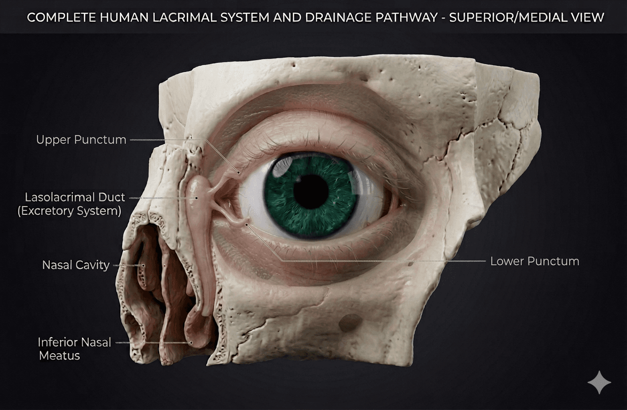
The lacrimal system produces, distributes, and drains tears. A healthy tear film is essential for corneal health, clear vision, and comfort. When any part of the drainage pathway is blocked, tears overflow onto the cheek (epiphora), stagnate in the drainage sac, and become a reservoir for infection.
For a detailed overview of lacrimal anatomy, see the Anatomy Overview or the dedicated Lacrimal Anatomy page.
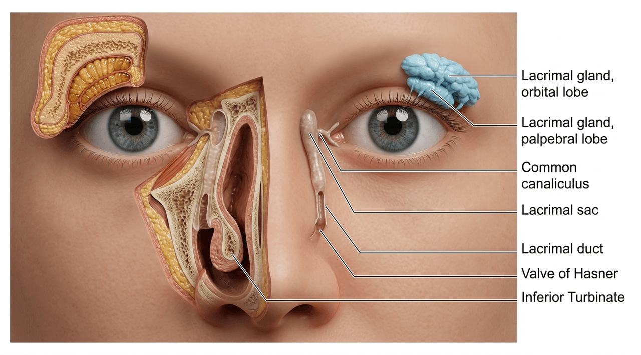
Understanding the anatomy guides both the diagnostic work-up and the choice of surgical repair. The drainage system has several potential sites of obstruction:
Tear-drainage problems range from a simply watery eye to blocked ducts, infections, and injuries. Explore each in depth:
Tears are made above the eye and drain through tiny openings (puncta) into the nose. When that drainage pathway is blocked, tears back up and symptoms appear:
Blockage can occur anywhere along the drainage path: age-related narrowing of the tear duct, chronic inflammation or infection, prior trauma or sinus surgery, canaliculitis (an infection of the tear channel), tumors, or — in babies — an incompletely opened duct (congenital tear-duct obstruction). Poor eyelid position or laxity can also cause tearing without a true blockage.
An oculoplastic surgeon distinguishes a drainage blockage from overproduction. See our Watery Eye Evaluation page for the full work-up, which may include irrigation and probing of the tear ducts, dye tests, and imaging to locate the level of obstruction.
Tear-drainage surgery is a core part of oculoplastic practice; an ASOPRS fellowship-trained surgeon can pinpoint the level of blockage and choose the right repair. Find one in our surgeon directory.
Related in-depth guides
Connect with a board-certified oculoplastic surgeon who specializes in lacrimal system.
Search the Directory →