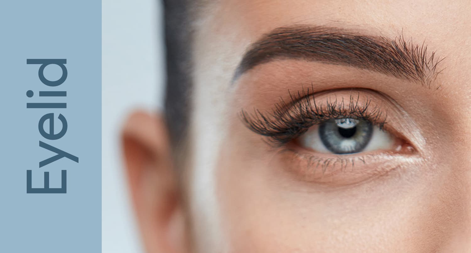
Ptosis
Repair of drooping upper eyelids (ptosis) — both cosmetic and functional correction of levator muscle weakness.
Medically reviewed by Mark S. Brown, MD — ASOPRS fellowship-trained oculoplastic surgeon
What Is Ptosis

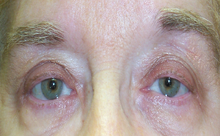
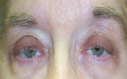
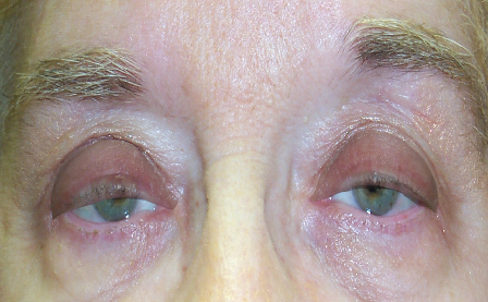
Ptosis (pronounced TOE-sis) is an abnormally low position of the upper eyelid. When the levator muscle — the primary eyelid elevator — or Müller’s muscle weakens, stretches, or detaches from the eyelid, the lid drops in front of the pupil. Ptosis may affect one eye or both and ranges from barely noticeable to severe enough to obstruct vision entirely.
Interactive: The Eyelid and Your Vision — Side by Side
Ptosis is not only a change in appearance — it directly removes vision. Use the simulator below: as you drag the slider, the upper eyelid on the left droops from its normal position into severe ptosis, while the panel on the right shows what the patient actually sees as the lid progressively blocks the upper (superior) field of vision. This superior field loss is what insurers measure with formal visual-field testing when determining whether ptosis repair is functional (covered) rather than cosmetic.
What insurers ask for (functional coverage)
- Visual-field test performed untaped and with the lid/skin taped up (showing improvement)
- Margin reflex distance (MRD) measurement
- Frontal and side photographs documenting the lid or skin position
- Documented symptoms (reading difficulty, brow fatigue, headaches)
Criteria vary by plan — confirm specifics with your surgeon's office.
Ptosis SimulatorEyelid Position & Visual Field


Drag the slider — as the eyelid droops, watch how much of the upper visual field disappears.
- Ptosis is also called blepharoptosis
- It differs from dermatochalasis (excess upper eyelid skin) — though both conditions frequently coexist
- A drooping lid that crosses the pupil reduces the superior visual field, causes eyebrow strain and headaches, and in children can lead to amblyopia (“lazy eye”)
For a detailed guide to levator anatomy, Müller’s muscle, and the tarsal plate, see our dedicated Anatomy Overview page.
Types & Causes of Ptosis
Ptosis is grouped by cause — age-related (aponeurotic), congenital, and neurologic. Recognizing the type guides both the work-up and the operation.
Explore Ptosis
Ptosis ranges from age-related drooping to conditions present from birth, each evaluated and treated differently. Explore each in depth:
Frequently Asked Questions
- What is ptosis?
- Ptosis (TOE-sis) is drooping of the upper eyelid caused by weakness or dysfunction of the levator muscle — the muscle responsible for lifting the upper eyelid. It can affect one or both eyes and may be present from birth (congenital) or develop over time (acquired).
- What is the difference between ptosis and blepharoplasty?
- Ptosis is caused by levator muscle weakness and requires surgical repair of the muscle itself. Blepharoplasty addresses excess skin overlying the eyelid. Both conditions can cause drooping or hooding, and they often occur together. Only an oculoplastic surgeon can reliably distinguish them and perform the correct procedure.
- How is ptosis repaired surgically?
- The most common technique is levator advancement — tightening the levator aponeurosis through an external incision in the eyelid crease. If levator function is poor (as in severe congenital ptosis), a frontalis sling procedure connects the eyelid to the brow muscle. Mild ptosis in patients who respond to phenylephrine drops can be corrected with a Müller's muscle-conjunctival resection (MMCR).
- Is ptosis surgery covered by insurance?
- Yes — ptosis repair is typically covered by health insurance when the drooping eyelid causes functional visual field obstruction, documented by a formal visual field test with the eyelid in its resting position.
- What should I expect during a ptosis consultation?
- During your consultation, your oculoplastic surgeon will perform a comprehensive eye examination, including measuring your eyelid height and assessing how well your levator muscle functions. You'll discuss your symptoms, review your medical history, and examine photographs to determine if you're a good candidate for surgery. The surgeon will explain the appropriate surgical technique for your specific condition and answer any questions about risks, recovery, and expected outcomes.
- What are the potential risks and complications of ptosis surgery?
- While ptosis repair is generally safe, potential complications include infection, bleeding, scarring, and asymmetry between the two eyelids. Some patients may experience temporary dry eyes, difficulty closing the eyelid completely, or under- or over-correction requiring revision surgery. These complications are uncommon, especially when performed by a fellowship-trained oculoplastic surgeon, and most resolve with appropriate care.
- What is the recovery timeline after ptosis surgery?
- Most patients can return to light activities within one to two weeks, though complete healing typically takes four to six weeks. During the first few days after surgery, you may experience swelling, bruising, and mild discomfort managed with prescribed medications and cold compresses. You'll have follow-up appointments to monitor healing, and your surgeon will provide specific instructions about activity restrictions, eye care, and when you can resume normal routines.
Find a Specialist
Connect with a board-certified oculoplastic surgeon who specializes in ptosis.
Search the Directory →